Welcome to our monthly newsletter, covering the latest large animal care advice and tips.‌ ‌ ‌ ‌ ‌ ‌ ‌ ‌ ‌ ‌ ‌ ‌ ‌ ‌ ‌ ‌ ‌ ‌ ‌ ‌ ‌ ‌ ‌ ‌ ‌ ‌ ‌ ‌ ‌ ‌ ‌ ‌ ‌ ‌ ‌ ‌ ‌ ‌ ‌ ‌ ‌ ‌ ‌ ‌ ‌ ‌ ‌ ‌ ‌ ‌ ‌ ‌ ‌ ‌ ‌ ‌ ‌ ‌ ‌ ‌ ‌ ‌ ‌ ‌ ‌ ‌ ‌ ‌ ‌ ‌ ‌ ‌ ‌ ‌ ‌ ‌ ‌ ‌ ‌ ‌ ‌ ‌ ‌ ‌ ‌ ‌ ‌ ‌ ‌ ‌ ‌ ‌ ‌ ‌ ‌ ‌ ‌ ‌ ‌ ‌ ‌ ‌
 
Orange Veterinary Hospital


57 Molong Rd
Orange, NSW, 2800
orangevet@orangevet.com.au


Large Animal News
February 2023
 

We've got a full compliment of vets back on deck, and the cattle pregnancy ultrasound back from servicing. 

The storms recently have seen plenty of horses finding themselves in fences, and ongoing good feed conditions are delivering plenty of fat, prolapsing beasts. 

 
Contact Us
 
Diseases of the mouth- Lumpy jaw & Wooden tongue 
 

Picture 1 (above): The common clinical sign shown by Cattle infected with Lumpy jaw.

A frequent cattle consult throughout the majority of the year is an animal that presents for swellings/ large lumps of the lower jaw. Most commonly the disease process going on here has been termed ‘Lumpy jaw’. Here there has been an insult to the inside of the mouth from fibrous material (rough grass/hay/sticks etc.) that have caused damage to the mucosa inside the mouth to which a bacteria called actinomyces thrives and invade. Over the coming weeks to months this area will become more infected, the infection then penetrates into deeper tissues, lymphatics and eventually the jawbone.

This is what vets call an ‘osteomyelitis’ (infection of the bone). As the disease progresses further, the bone is eaten away by the infection and the animal will eventually lose its ability to chew and therefore lose condition. In most cases have to be euthanised.

The key to avoiding this undesirable outcome is to act promptly once any clinical signs develop. Like any disease the sooner a producer gets a veterinarian involved, the sooner the issue can be fixed. If this disease if left untreated the animal will suffer and reach a point where the disease is untreatable.

Moral of the story; a thorough examination of all animals frequently even just in the paddock will most likely avoid this disease developing to a point where it cannot be treated. Here we would love producers to be looking for animals with swellings under the jaw, excessive saliva coming from the mouth and even a loss of condition.

Picture 2: Top; Normal jaw bone. The bottom of the picture shows the jawbone of the cow that has been infected with ‘lumpy jaw’.

Wooden tongue is a lay term for the disease called ‘Actinobacillosis’. It like Lumpy Jaw is a disease of the oral cavity. Like the name suggests, the tongue becomes like wood (hardens). Much like the lumpy jaw information previously mentioned, as the disease progresses the animal loses the ability to chew and swallow; leading to the animal losing condition and eventually dying or having to be euthanised.

The common ground that these two diseases share is that they are caused by bacteria from the same family. The most common clinical sign to look for is a cow with excessive saliva production, swelling under the jaw and the tongue slightly protruding from the mouth. The reason for this is that the tongue swells significantly as the disease progresses. In terms of treatment if the diseased animal is identified early in the disease process it can be successfully treated. Don’t wait to ‘See how it goes’.
The last point to make is that this disease can also affect sheep. In sheep however it is more common for them to develop lesions on their lips rather than their tongues.

Picture 3: Common clinical sign displayed by cattle with wooden tongue.

 
Alpacas- unique critters, unique needs 
 

Alpacas can be wonderful pets, brilliant gaurdian animals and produce fantastic fibres, but their unique looks are just one thing that makes them special. As part of the camelid family, their health needs are closest to the camel, not the sheep.

Alpacas originate in South America- a far cry from the fertile soils in the Orange district. Raising, farming and caring for alpacas is a bit different to their lives in South America. 

Keeping alpacas healthy locally often requires a bit more work than you might be bargaining on: 

- desexing males helps population control, and fighting between males

- alpacas are non-shedding, so summer shearing is a must 

- foot care with alpaca farriery might be needed, usually at shearing

- they can be escape artists! Mesh fences help with keeping families together

- alpacas make great gaurdians- keep this in mind with dogs around

- parasite problems can emerge- faecal monitoring helps plan drenching 

- vaccinations are vital- a '5in1' or '7in1' vaccine might be used 

- Vitamin D can be seasonally neccessary - injections are best 

- alpacas need lots of roughage- plan on 1-2kg daily (grass + hay are best)

- alpacas need fresh water available- plan for up to 4L a day per animal

- if you have alpacas, you should have a registered PIC with the LLS

- teeth can be troublesome, especially if there is a bite issue. Check each year

 
Worming guide for alpacas, goats and sheep
 
The case for heavy heifers - a major profit driver!
 

Heifer rearing often accounts for a big chunk of the input costs of running a farm every year, but did you know that an investment in rearing healthy heifers is an investment in the future productivity of your farm?

So how can farmers get the best out of first calving whilst maintaining a healthy, productive and fertile heifer?

The age of a heifer, coupled with their rate of growth, weight and frame size at first calving are the major variables that contribute to their health, productivity and fertility.

Well-grown heifers are also more fertile at their initial joining, and throughout subsequent breeding once in the herd.

 
Bush ticks – the transmitter of theileriosis
 
Bush ticks have received plenty of attention in recent times due to their role in the transmission of the disease ‘theileriosis’. Most infestations are relatively light and/or short-lived, however, the introduction of tick-naïve cattle into tick-endemic areas can lead to heavy infestations with serious consequences. In these instances, “tick worry” causes anaemia among the herd, as well as significantly reducing cattle live-weight gain and milk production.

Where do bush ticks live?
The bush tick is found mostly in sub-tropical regions and some temperate areas with summer rainfall. While they prefer to live on cattle, they’ll also reside on deer, sheep, pigs, dogs, horses and even humans. They like warm weather and plenty of rain and are generally found on the areas of the animal that are hard to groom, like the brisket, udder and inguinal regions, and in the ears. Fortunately, ticks are not a big problem locally but a good reason to review your biosecurity and quarantine practices with introducing new stock. 

Can we control a bush tick infestation?
Controlling bush and paralysis ticks is difficult because:
  • They are multi-host ticks and only stay on an animal for a week at a time.
  • They also infect native animals (i.e. reservoir hosts, eg kangaroos).
  • They can survive for up to 9 months on the ground without a host
  • Current spray chemicals are short-acting, and are both slow and labour-intensive to apply.
Understandably, bush tick management is a source of frustration for many farmers. That said, Tickboss provides great information and resources on the control and treatment of bush ticks. You can also view this webinar on ticks and theileriosis management.
 
Managing theileriosis outbreaks on Australian cattle farms
 

Theileriosis is a disease affecting cattle that has caused significant economic losses for many Australian farms. First appearing in New South Wales in 2006, the disease then spread to Victoria (mainly Gippsland) in 2011, and has just been detected for the first time in Tasmania. Historically, it has been reported in many parts of NSW, including the Central Tablelands (click on the cows above to see a map of historical cases).

What causes theileriosis?
The disease is caused by the protozoan parasite (Theileria orientalis ikeda). Signs of the disease are similar to symptoms of severe anaemia and may include:

  • Lethargy
  • Poor appetite
  • Exercise intolerance
  • Increased respiratory rate
  • Pale to jaundiced mucous membranes, often giving a pale and yellowish appearance to the eyelid
  • High temperature
  • Abortions
  • Drop in milk production in dairy cows
  • Death, most commonly in heavily pregnant cows

How to diagnose theileriosis?
Theileriosis can be easily diagnosed with a blood test.

How does theileriosis spread?
The bush tick (Haemaphysalis longicornis) is the usual vector that spreads theileria. Other carriers such as biting flies, sucking lice and vaccination needles are also capable of spreading the disease.

How is theileriosis treated?
Several medications are available to treat theileriosis, depending on the severity of the disease. If cattle are severely anaemic, blood transfusions may be necessary, and the prognosis deteriorates significantly.

How can you protect your farm against theileriosis?

  • Avoid introducing cattle from affected areas
  • If you must introduce cattle:
  • Introduce from districts where infection is uncommon
  • Perform biosecurity procedures throughout the introduction:
  • Treat for ticks on arrival
  • Place in paddocks less favourable for ticks
  • Separate introduced cattle from home cattle
 
Leg wound care for horses
 

In summer, it seems that less time spent munching grass or going to competitions means more time for mischief and misadventure. This combined with some horses’ nervous, flighty disposition can result in bumps, grazes and sometimes cuts.

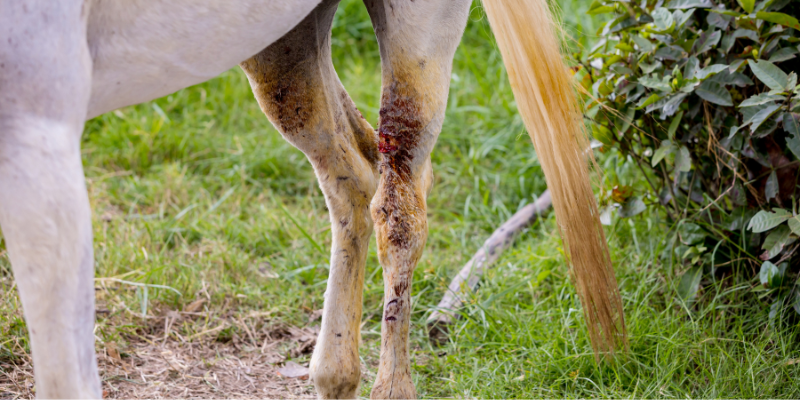
When a cut occurs on a part of the horse which is well covered with muscle or other soft tissue, complications with healing after veterinary treatment are not common. However, if a significant injury breaks the skin on the lower part of the leg, then the chance of complications with healing increases significantly.

There are two main reasons for this:

  1. The lower leg of a horse has much less muscle under the skin, mostly just bone, tendons and joints. This means when skin is cut and shrinks from the wound site, as skin tends to, stretching the skin so it joins normally is more difficult. This is because muscle under the skin is more flexible than bone and tendons.
  2. This area has a lower blood supply as bones and tendons need relatively little blood compared to muscle. This blood supply is closer to the skin surface and easily damaged when horses are injured.
    So, the combined difficulty getting the skin to join and getting optimum blood supply for good healing makes for a challenging recovery for the horse, carer and vet.
What first aid can you apply?
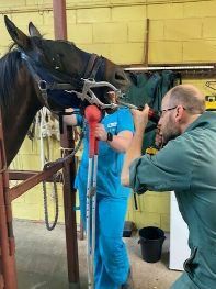
Apart from your vet’s phone number, it is important to have a plentiful supply of wound protection on hand. After identifying a cut or graze, your first priority is to stop the bleeding. Next, clean and cover the wound. For this, you’ll need some sterile gauze which won’t stick to the wound. Then, it's necessary to apply some good disinfectant like Betadine or Hibitane. You will need to protect the wound with some gauze-covered cotton wool roll, which you can hold in place with good-quality elastic bandages. Once the wound is well bandaged, get the horse in a clean safe area, while you wait for your vet to arrive. While you wait, it's a good idea to recall your horse’s tetanus vaccination history. Horses are more sensitive to tetanus through wounds than any other species.

When is veterinary care required?
The initial treatment of a wound, particularly of the lower leg can have a profound effect on the long-term outcome for your horse.
 
A vet is urgently required when:
  • The wound involves a blood vessel or a joint
  • The wound edges won’t easily join
  • The wound is contaminated with dirt or other material
  • You can see underlying structures such as joints or 'white bits'
  • The horse is lame as well as a laceration
  • The main issues your vet can address quickly are stopping blood loss, infection management, and strategies to improve wound healing, which can include suturing, skin flaps and/or grafts to fill gaps in skin coverage.

Post-Surgical Management
The management and care of these sometimes-complicated wounds is often a shared responsibility between horse, carer and vet. Vets need skills which go far beyond surgical skills. A vet and the owner will team up and discuss so that they can decide what level of responsibility the owner can safely undertake to change dressings, assess how the wound is healing and redress the wound.

The vet will then decide how much instruction to give the owner verbally and in writing to ensure these important tasks are carried out effectively. The vet can then decide how often they need to visit the horse to check and re-bandage the wound.

Sometimes blood supply to the healing wound is compromised and the outcome is not ideal. For example, a poorly healing wound may result in surrounding skin dying, which then necessitates a secondary healing process such as a red area. The formation of a red area, sometimes called granulation, involves covering the underlying structures with tissue that is rich in blood vessels until the skin heals from the outside in. Although not ideal aesthetically, the red area will ultimately be covered with skin scar tissue. This scar tissue is not as flexible and may restrict some movement, so this treatment should be avoided if at all possible.

Your vet, diligence and patience are your best friends when it comes to complex wound healing.

 
Castration laws – what farmers can and can’t do, and how to do it humanely
 

Calves should be castrated as young in life as possible to reduce blood loss and pain. We strongly recommend providing pain relief whenever possible during castration- it helps the (soon to be) steers recover faster. 

Here are a few guidelines you should follow when castrating your calves:

Standards (legal requirements which must be adhered to)

  • The person in charge of castrating must ensure the use of appropriate pain relief when castrating cattle, unless the cattle are:
  • less than six months old, or
  • less than 12 months old when at their first yarding and where the later age is approved in the jurisdiction.

Guidelines (non-compulsory but strongly recommended practises)

  • Castration should be completed as young as possible (less than 12 weeks of age) and prior to weaning.
  • Calves should be more than 24 hours old when castrated.
  • Calves less than two weeks old should be castrated by the rubber-ring method.
  • Rubber rings or tension bands should be correctly positioned to ensure tension is achieved to block the arterial blood flow. This should only be applied when two testes can be palpated in the scrotum beneath the rubber ring
  • Calves older than two weeks old should be castrated by excision which extends fully to the base of the scrotum to allow effective drainage and reduce the risk of infection.
  • Surgical procedures should be done with pain relief. Operators should seek advice on current pain minimisation strategies.

For further advice, or to plan an upcoming castration, speak to a member of our knowledgeable veterinary team. If things don't seem right, you can get one of vets to assess and make a plan together. 

 
Q-fever – new research indicates all rural residents are at risk
 

New research has been published suggesting that rural residents should be vaccinated against Q-fever. Q-fever is a bacterial disease transmitted to humans primarily from cattle, and other ruminant animals like goats and sheep. The disease can have a lifelong impact on health, including post-Q-fever fatigue syndrome. Most of the time, an animal infected with Q-fever shows no symptoms, however it may cause animals to go off their feed, and is known to cause late-term abortions.

The study found one-in-20 people in rural Queensland have been exposed to the bacteria that causes Q-fever, and a rate of one-in-27 people in rural New South Wales. Interestingly, this study still is likely to underestimate the true exposure rates as antibodies (which indicate exposure) decline below the limit of detection with time. The single-shot vaccine is available for humans (not animals), and is highly-effective at conferring lifelong immunity against the disease.

In light of this information, vaccination (or testing at the very least) is recommended for all people with frequent exposure to sheep, cattle and goats, as well as abattoir workers and farm animal birthing assistants, and to any resident of a rural area who is likely to come into contact with somebody exposed to Q-fever. Usually the first step of Q-fever vaccination are tests to check that the person has not been exposed to Q-fever already.

Information about Q-fever and finding medical centres that vaccinate against it are available on the Australian Q-fever register.

 
Equine Odontoclastic Tooth Resorption and Hypercementosis
 
Equine Odontoclastic Tooth Resorption and Hypercementosis (EOTRH) is an uncommon disease which results in the resorption (the loss or partial loss) of incisors and occasionally other teeth in older horses. It has only recently been described as a condition in its own right, distinct from the resorption of teeth in horses over 15 years old, which is relatively common.
 
EOTRH begins with subtle signs that gradually progress until quite severe, often before diagnosis.
 
Early signs can include:
  • Inflammation and recession or overgrowth of the gums
  • The area below the gums may be swollen and teeth can show a bulbous enlargement
  • Pain responses, including misbehaving or changed behaviour
Progressive signs can include:
  • Periodontal disease
  • Pathologic tooth fracturing
  • Tooth looseness before complete tooth loss
Diagnosis
X-ray of the incisors is usually required to diagnose this disease. These x-rays will determine how many teeth are affected, and how severe the lesions are.
 
Causes
The cause for this syndrome is not known, however theories include physical stress associated with the biology and use of the horse’s mouth, as well as secondary bacterial involvement.

The course of the disease
As EOTRH progresses, the roots of the incisors (and sometimes canines and cheek teeth) undergo an inflammatory process and begin to dissolve. This happens often in combination with excessive cemental deposition. This hypercementosis is considered an attempt by the body to repair the teeth rather than a primary disease process. This over-deposition of tooth cement appears as bulb-like swellings around the roots of the affected teeth.

Sadly, with disease progression, affected teeth gradually become increasingly loose and painful. Serious resorption can result in teeth fracturing, as well as infections as the roots dissolve. These infections appear as small red dots above the gum line of the incisors, or even small pimple-like draining tracts in the gum area.

Often older horses are pretty tough and will not show obvious pain until the disease is well progressed. As the disease begins to affect eating ability, horses can lose weight and be reluctant to bite down on treats or carrots, and stop grazing due to incisor discomfort.

As pain increases horses may become sensitive to being bridled or even ridden.

Treatment
Because the cause is not understood, there is no known current treatment or preventive action for EOTRH, apart from extraction based on the severity of the lesions (as shown by an x-ray). Depending on the skills, tools and experience needed, there are equine veterinary dental specialists that may need to be consulted. 

Recognising the early signs of the disease may enable more effective symptom management, and due to the progressive nature of the disease, increasing pain is often the most serious concern for horses affected by EOTRH.
 
For that reason, strategic tooth extraction can have an improved prognosis and is helpful in managing long-term outcomes.
 

This email contains comments of a general nature only and is not intended to be a substitute for professional veterinary advice. It should not be relied on as the basis for whether you do or don't do anything. 

All content © Pet Pack 2022

This email was sent to: email@example.com

Orange Veterinary Hospital
57 Molong Rd
Orange, NSW 2800